![]()
Color Trading Sp. z o. o. Philippines site
- English
Please select your Region.
Please select your Region.
![]()
Color Trading Sp. z o. o. Philippines site
Unique Features
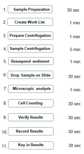
Manual Microscopy
- Approx. 10 - 12 minutes per sample
- Takes too much time of "highly qualified" lab personnel
- Sample preparation
- Slide preparation
- Slide viewing
- Abnormal findings are always questionable
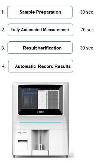
Automated Urine Microscopy
- Approx. 1 ½ minutes per sample
- > 25000 images of patient's results can be stored & shared
- Possible to review and verify results
- Process security
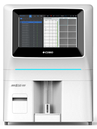
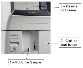
S50 - Simple to Run Samples
1. Put urine sample at sample holder
2. Click on start button
3. Results on the screen along with microscopic images
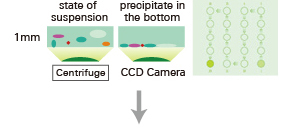
S50 - Instrument Measuring Sequence
Centrifugation is performed to create a monolayer of particles at bottom of the cuvette
Used cuvette is placed into waste-bin
S50 - Instrument Measuring Sequence
sample injection into cuvette
accelerated sedimentation, auto-focusing
AI automatic recognition
iClinical.net
Measurement Technology
- Sample preparation
- Sedimentation
- Microscopy : focusing, image capturing
- Particle recognition
- Particle classification
- Particle labelling
- Summarizing the detected particles
- Unit conversion
- Semi-quantitative categorization
Measurement Technology - Sample Preparation
- S50 is equipped with in-built barcode reader
- Bi-directional LIS / HIS
- Mix urine before aspiration
- Probe wash after each transfer of liquid
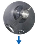
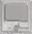
Measurement Technology - Patented Cuvette
- Each sample is analyzed in a separate cuvette
- No carry over between samples
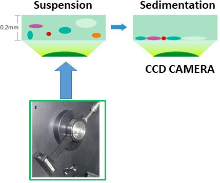
Measurement Technology - Sedimentation
- Centrifugation @ 2000 RPM, for 10 seconds
- Create monolayer of particles at the bottom of the cuvette
- Drive all particles to the same level to achieve good focusing
- Better pictures based on good focusing
Measurement Technology - Microscopy
- Multiple digital images are taken by CCD
- Possible to chose 5, 10, 15 or 20 images per sample (default setting is 15)
- Images and results are displayed, rechecked as well as corrected
Measurement Technology - Artificial Intelligence
- S50 works on deep learning algorithm
- Algorithm of S50 need 3000 marked pictures
- Take pictures of thousands of samples
- Marked the name of particle in picture by morphologist, i.e. RBC, WBC . . .
- Different cells have different physical properties
・ Shape, Size, Diameter & Thickness
- It takes these physical properties, models them, and trains them to forms algorithm of S50

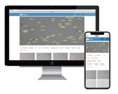
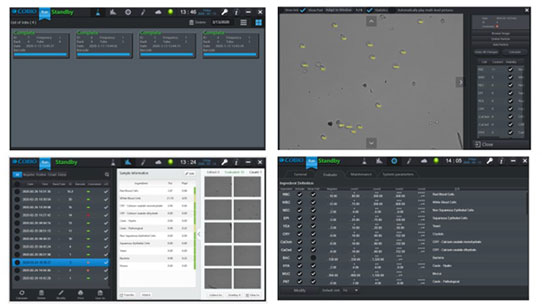
Result Display - Screen Shots
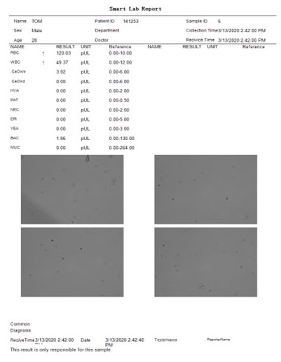
Report Printing
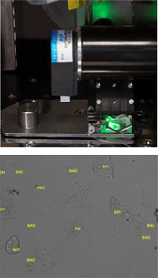
Automatic Label
RBC, WBC, HYA, PAT, EPI, NEC, BAC, YEA, CRY, CaOxm, CaOxd, MUC
Manual Label
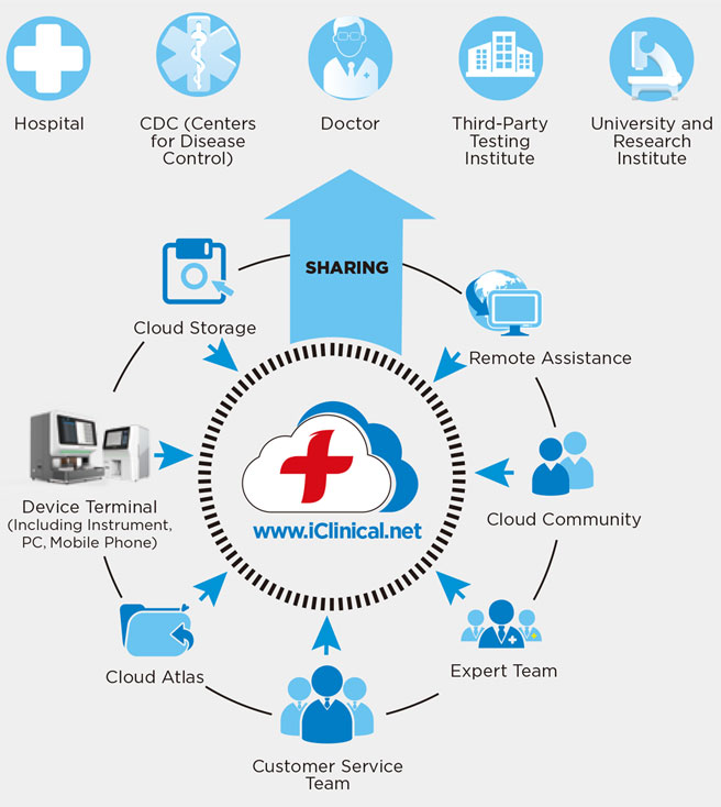
iCLINICAL Cloud Service
- Online training
- Online help
- Online sharing
- Knowledge management
- Access to historical data analysis and research
Test Performance Data
- Coincidence rate of identification (versus manual microscopy)
- Red Blood Cell ≥ 98%, White Blood Cell ≥ 96%, Tubular ≥ 72%
- The false negative rate of analyzer (versus manual microscopy) is zero
- When the cell concentration is 50 / uL, CV ≤ 18%
- When the cell concentration is 200 / uL, CV ≤ 11%
- CV of total cell count results is ≤ 14%
Test Performance Data
- The analyzer's blank count for WBC and RBC is zero
- S50 analyzer can detect RBC / WBC samples with concentration level of 5 / ul
S50 - Salient Features
![]()